

Medical imaging and medical image analysis represent the core of digital health. Evaluations based on Deep Learning algorithms are now employed to solve cases that other technologies cannot cover. In his lecture, Anders Heyden from Lund University, Sweden, gives an eloquent and complete definition of medical image analysis. He says:
Medical image analysis is the science of solving/analyzing medical problems based on different imaging modalities and digital image analysis techniques.
In this article, you will learn about the following:
- Types of medical image analysis
- Medical image analysis: Bacteria counting
- Magnetic Resonance Imaging, MRIs
- Mammography 3D image analysis
- 6 Steps how AI performs medical image analysis
- AI libraries for medical image analysis
AI for Medical Image Analysis Business Case
And if you are looking for medical image analysis software development services, don’t hesitate to contact us and learn more about our expertise in this field.
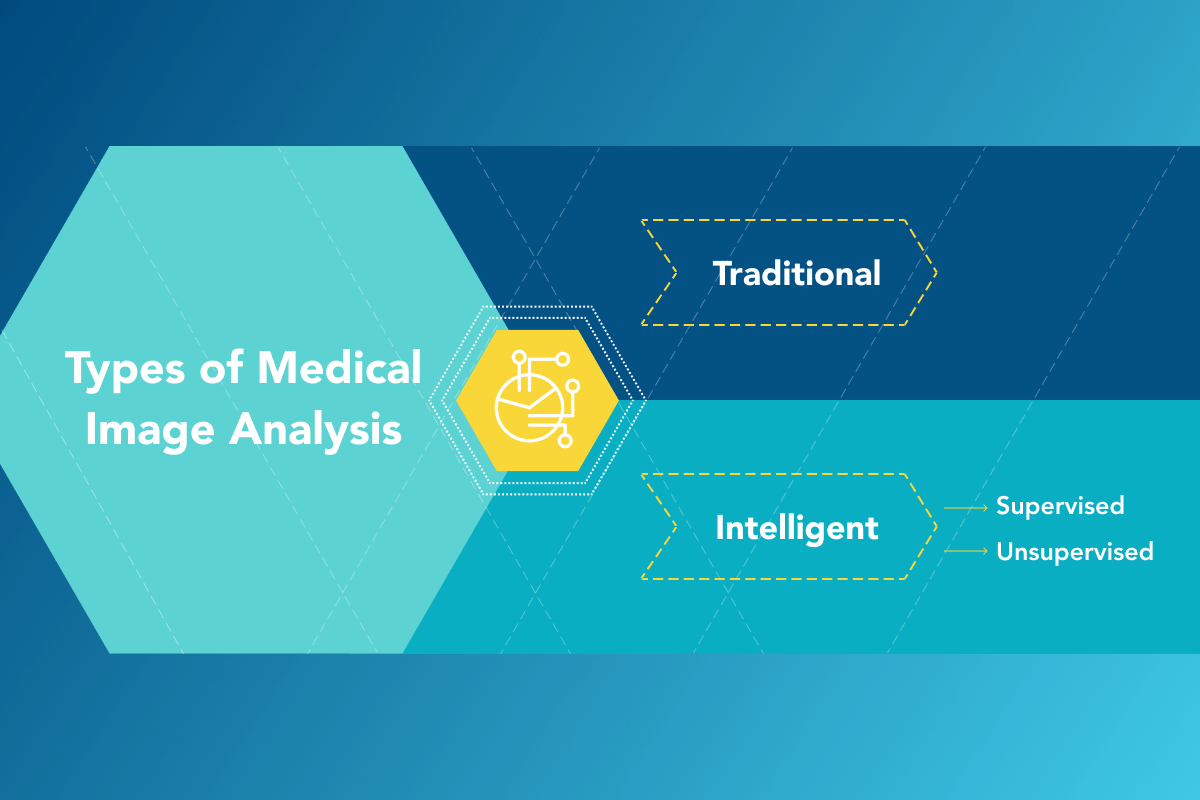
Types of Medical Image Analysis
1. Traditional medical image analysis is applied to small volumes or almost similar-looking objects with similar features.
2. Intelligent medical image analysis uses Computer Vision and Machine Learning and is applied to large volumes or not-matching to each other objects. Intelligent medical image analysis can be subdivided into:
- Supervised
- Unsupervised
Intelligent solutions are effective in complex or massive tasks and can be retrained for different types of such tasks.
Applications of Medical Image Analysis
The application of AI for medical images analysis is very wide and can include:
- Endocrinology (to detect diabetes);
- Oncology (all kinds);
- Ophthalmology;
- Surgery assistance;
- Tasks in medical laboratories;
- Predictive diagnosis and more.
Solutions for medical image analysis can work in different modalities: mammography to predict breast cancer, chest CT and Magnetic Resonance Imaging MRI, lung cancer X-ray, ultrasound, and neuroimaging. Thus, it can support common medical image formats, such as DICOM (3D images, Digital Imaging and Communications in Medicine), AMINC, ECAT7, NIfTI, and more.
Benefits of Medical Image Analysis
The benefits of medical image analysis are indeed hard to count and depend on applications and modalities. But they can be generalized. Due to intelligent medical image analysis, doctors and researchers can:
- Save time and attention with routine and big data volumes;
- Define precise segments for medical treatment;
- Detect the slightest abnormalities and lesions in the early stages.
Medical Image Analysis: Bacteria Counting
The research of Cornell University, USA submitted March 2021 considers:
Microorganism counting techniques are crucial in microorganism analysis helping biologists <…> calculate their biomass concentration and biological activity.
The research summarizes image segmentation methods by periods in 142 papers in a single endeavor to find trends in microorganism counting.
Deep Learning Trends in Bacteria Counting
In bacteria counting, scientists adhere to edge trends and technologies such as Deep Learning in combination with traditional plate methods. Thus, Oxford Academic first offers its year 2020 detailed review based on Machine Learning, data science, biofilm, segmentation, and phenotyping.
<…> how bacterial cells and communities can be detected in images using traditional image analysis and convolutional neural networks, and how quantitative properties can be extracted after the object detection.
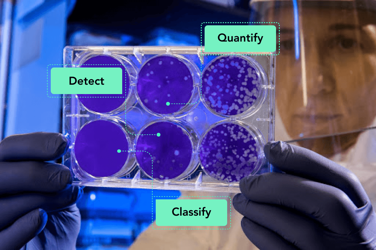
Picture of The Public Health Image Library from the Centers for Disease Control and Prevention (CDC)
On the picture:
The scientist examines the result of a plaque assay, which is a test that allows scientists to count how many flu virus particles (virions) are in a mixture.
Softengi – AI solutions developer company – is successfully extracting quantitative properties from medical images of bacteria communities, having trained the model that can detect bacteria with 99% accuracy. This means that with this solution, you can count 99% of all bacteria in the image of the plate and be absolutely sure about their concentration and biological activity. The solution detects, classifies, and counts bacteria using Faster RCNN and Resnet101 neural networks, TensorFlow, Open CV, and Keras.
Softengi and Supervised Bacteria Counting
To count bacteria, Softengi trained or supervised machine learning to detect and classify bacteria. Machine learning performs detection and classification based on an AI library with thousands of labeled bacteria images, characteristics of bacteria, and functions. Creating such a library is rather long work for developers. Thus, the resulting solution can recognize many objects in medical images and do it promptly.
Softengi and Unsupervised Bacteria Counting
Softengi also develops unsupervised learning and convolutional neural networks for medical image analysis. Unsupervised learning can learn patterns from unlabeled data, and clients can train it with their own libraries and goals. This makes unsupervised learning not only effective but also a very scalable solution for medical image analysis.
Unconditionally, both supervised and unsupervised machine learning solutions for bacteria analysis make the lives of doctors, patients, and lab personnel better.
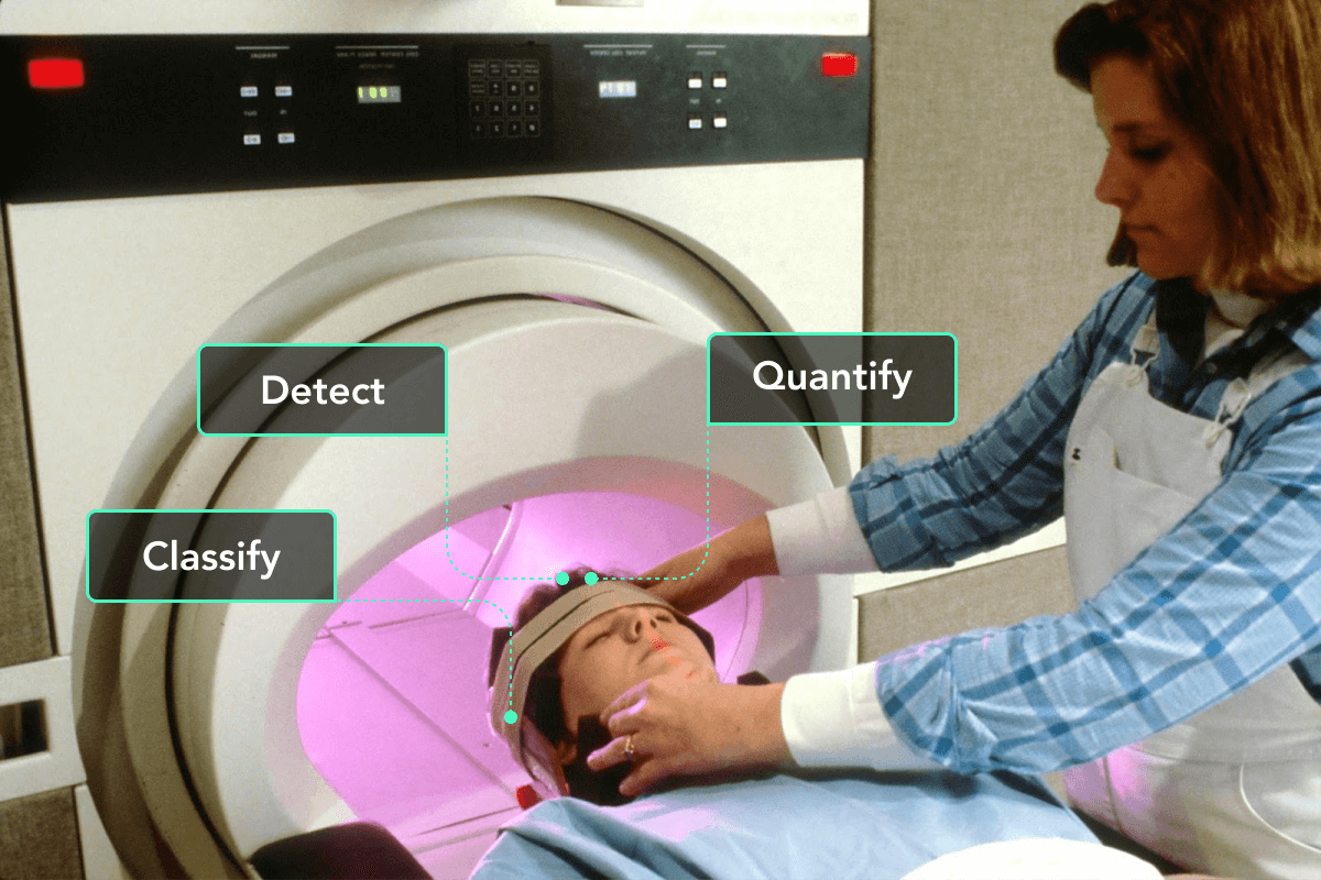
Magnetic Resonance Imaging, MRIs
Doctors request an MRI to detect diseases of the inner organs of the body as tissues. MRI scanners apply magnetic fields and radio waves to make images of the organs in the body. MRIs require zero radiation, which makes them very popular with doctors and patients. Artificial Intelligence solutions in MRIs can:
- Significantly reduce the workload on radiologists;
- Analyze numerous complex scans using detection filters and mathematical methods;
- Propose edge approaches in detecting and classifying hard diseases.
Now, look at Deep Learning trends in MRI and Machine Learning applications in cell image analysis.
In classifying Alzheimer’s disease, the new Deep Learning-based chromosome encoding approach proposes applying a Genetic algorithm to the brain MRI scans. This algorithm has extracted 6 to 65 knowledgeable brain cells in a segment of the brain that 3D Convolutional neural networks usually use to classify brain diseases. This means that Alzheimer’s disease, which is still on hold to be cured, can be detected in early stages using Machine Learning in MRI.
Another Deep Learning medical image analysis software can promptly classify different pathologies of knees on MRI scans. It can use internal and external libraries that make it easy to teach new knee abnormalities. This software can help doctors analyze MRI scans of keens and make correct diagnoses for sportspeople, seniors, and injured in accidents.
Mammography 3D Image Analysis
From 2 to 7 breast cancers are detected per 1000 women via mammography, says Summary of Cancer detection rates.
3D mammography is considered to be more accurate than 2D, and that is why more women choose the 1st method. Though, it costs a little more and takes a bit longer to do and to read. Also, insurance may or may not cover mammography. Still, with 3D mammography, 15% fewer patients are asked for additional evaluation.
Computer Vision and Machine Learning solutions for mammography analysis can promptly recognize benign and malignant tissues and detect other abnormal formations.
“In a study published in Clinical Cancer Research, deep learning was used to analyse a large set of mammograms to distinguish images with malignant diagnosis from benign lesions <…>. Deep learning distinguished recalled-benign mammograms from malignant and negative images. This study showed that there are imaging features unique to recalled-benign images that deep learning can identify to help radiologists in decision making to help reduce unnecessary recalls.” – reports Healthmanagement Volume 19.
The intent of mammography is to find abnormal non-fatty breast formations in a number of images of the same breast.
Deep Learning Trends in Mammography
A very accurate software with neural networks for mammography image analysis uses images taken from different angles and can detect the smallest cancer cells in mammography images with radiological accuracy.
Another mammography image “quality assurance” neural network has been trained to predict image quality based on a single mammography image that can illuminate a long pre-processing phase.
These and many more High Technology methods raise the level of breast cancer detection, do not expose patients to radiation, and cut the time of mammography procedure. Edge Deep Learning trends in medical investigation continue to move forward in lesion detection and computer-aided decision-making.
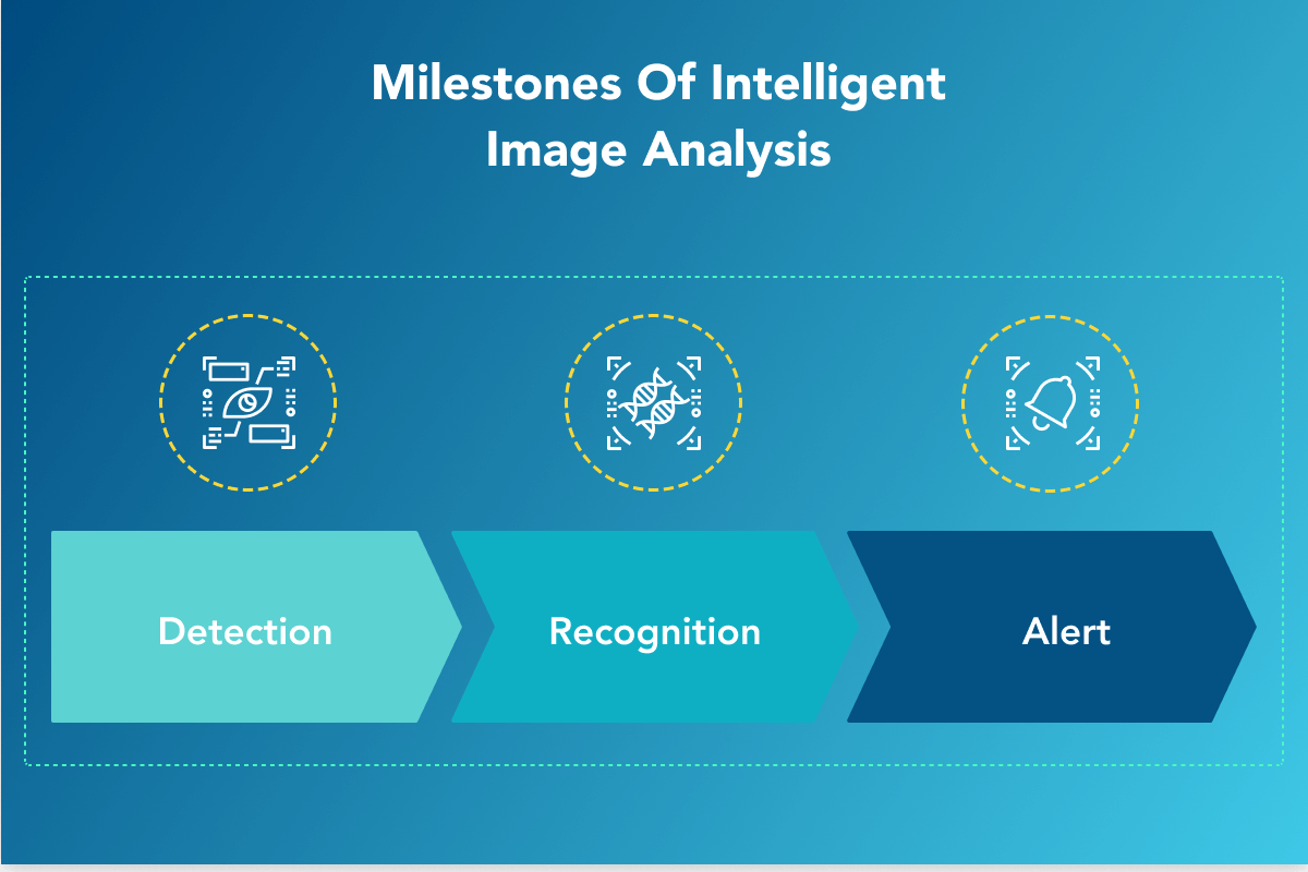
6 Steps How AI Performs Medical Image Analysis
Steps of Intelligent medical image analysis can be generalized into 3 milestones similar to all Machine Learning solutions:
- Detection;
- Recognition;
- Alert.
Depending on the task, Deep Learning medical image analysis software can perform 6 steps:
- Image processing;
- Segmentation;
- Detection;
- Classification;
- Registration;
- Quantification.
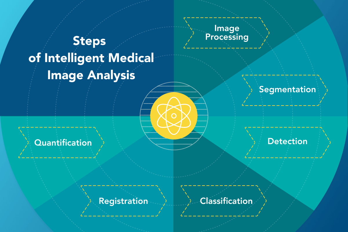
The quality of images is important to detect the requested objects, and developer companies can process the original images if needed. To identify the abnormalities, the solution segments all the objects on the medical image. Then, the solution detects and classifies the objects using the library. Overlapping or registering images taken in different modalities (from different diagnostic equipment) or images taken in different periods in dynamics is a very important step in image analysis AI medical field for doctors’ decision-making. The detected and recognized formations can be further quantified in size, form, and structure. With these steps, intelligent medical image analysis solutions aim to analyze complex and numerous images quickly and scrupulously.
To run through these steps, medical image analysis software must be well-trained and operate with huge libraries.
AI Libraries For Medical Image Analysis: OpenCV
There are several libraries that AI can use to perform effective deep learning for medical image analysis as OpenCV for image processing and computer vision, TensoFlow to construct and train neural networks targeted to find and classify images, PyTorch to accelerate the launch of solutions in industrial scales, Caffe a machine learning library for precise segmentation of images, VCXL for C++ and many others.
For example, OpenCV is a popular open-source library with programming functions for real-time Computer Vision and ML. It can be used for object detection, image segmentation, optical flow, face recognition, input/output of images, data structures, continuous integration, and is cross-platform.
Developer companies choose the most fitting AI libraries for each solution to perform deep learning in medical image analysis most effectively.
Functions of Computer Vision and Deep Learning
Using AI libraries, Computer Vision and Deep Learning can perform various tasks with objects on videos and images, including:
- Raising resolution on images with low resolution;
- Localization of abnormal cells on medical images;
- Classification of bacteria on the images of a plate;
- Segmenting while identifying certain pixels that belong to objects;
- Assembling when stitching segments for better field of view (FOV) or overlapping images of the same objects made in dynamics;
- Coloring to outline unhealthy formations on medical images;
- Recreating missing parts of objects on images with flaws;
- Detecting objects with red boxing them on videos.
Cross-Industry Applications of Computer Vision and Deep Learning
Applications of Computer Vision with Deep Learning go far beyond medical image analysis and can include: security and surveillance, traffic control, agriculture, manufacturing, and many more. Unsupervised learning can provide customers with solutions that can be retrained for different purposes with different objects and use AI libraries in various applications, including medical image analysis.
Conclusion
Computer Vision and Machine Learning are being vigorously employed in medical image analysis for better detecting of lesions on bodies and images. Deep Learning medical image analysis software has many applications and works with many modalities. The main purpose of Deep Learning for medical image analysis is to unload medical personnel from routine work with large data (bacteria counting), recognize large amounts of diverse data (multiple lesions), and recognize single small deviations from the norm. To do that, AI developer companies use supervised and unsupervised Deep Learning in medical image analysis. Edge Deep Learning trends in MRI and mammography give way to better detection of hard diseases such as Alzheimer’s disease and breast cancer. Scientists and developer companies work hard to design and train effective Deep Learning solutions with extensive libraries to maintain the highest image analysis standards in healthcare, make them affordable, and raise the quality of life.